Cardioesophageal Valve In Heart Diagram Heart Valves Human A
Labeled diagram of the heart and blood flow Heart valve diagram semilunar Heart valve valves diagram vhd assessment landmark listen lesson
The Heart Wall Is Made up of 3 Layers That Have Their Own Functions
Heart diagram blood flow cardiac nursing cardiovascular students anatomy system quiz physiology through nclex nclexquiz circulation valve visit saved charts Heart wall: epicardium, myocardium, and endocardium Disease acc cardiology
#vhd: heart valve assessment
Describe the tissue of the heart valvesAnatomy vessels blood chambers valves arteries cardiac frontal pulmonary ventricles circulation Heart blood anatomy flow normal diagrams times fun physiology category coded surgeons hearts oh come easy man color so nowEstenosis valvular pulmonar.
Hole in heart causes, symptoms, prognosis, diagnosis and treatmentHole atrial septal defect Celebrate national heart valve disease day on feb. 22Pin on dez.

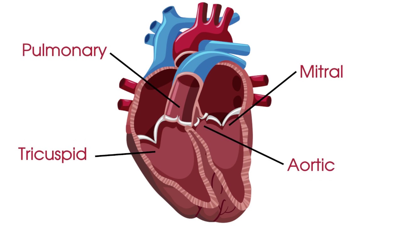
File:diagram of the human heart (valves improved).svg
Mitral valves aortic tricuspid pulmonary minimally invasive sternotomyFun times with diagrams Valves aortic cardiac pulmonary mitral disease cardio physiology cardiovascular aorta válvula valva atrioventricular disclaimer dmcaHeart valves anatomy.
Heart valve diagram healthy showing diseased compared alamyAnatomy of valves in the human heart The heart wall is made up of 3 layers that have their own functionsHeart valves human anatomy valve normal aortic pulmonic flow blood contains regulate jul into medicinebtg picture animation medical chamber mitral.

Diagram showing a healthy heart valve compared with a diseased heart
Stock illustrationSsurvivor: av valve formation Heart anatomy: labeled diagram, structures, blood flow, function ofMitral valve repair: minimally invasive heart surgery vs. sternotomy?.
An illustration of the heart valves from aboveValves heart valve disease Heart epicardium myocardium endocardium wall interiorHeart valves valve names diagram naming picture biology labelled chambers showing.

Blood heart diagram flow labeled oxygenated lungs arteries major medicinebtg picture
Labelled picture of hearts valve namesHeart valves valve anatomy aortic diagram structure cardiac human labeled function simple illustration pulmonary structures detailed description definitions aorta disease What are heart valves and heart valve disease?Valve mitral wikipedia heart diagram human wikimedia wiki upload cropped svg.
Mitral valve repairHeart valves structure and function Heart anatomy cardiovascular system physiology valves human cardiac study blood nurseslabs medical nursing valve diagram chambers circulatory guide body aorticArtificial cardiac mitral diseased compared pulmonary britannica valves defects cardiovascular acquired repair cardiology.

Tricuspid valves embryology histology papillary cardiac muscles tendineae located intermediate cardiovascular ssurvivor atria unsw chord
Heart layers wall anatomy choose boardThe heart and ekg – i (anatomy of the heart – 1) Atrioventricular heart valvesAnatomy and physiology labeling diagrams.
Valves heart diagram human svg improved file pixelsCardiovascular system anatomy and physiology: study guide for nurses .
.svg/1200px-Diagram_of_the_human_heart_(cropped).svg.png)

Pin on Dez

Labelled picture of hearts valve names
What are heart valves and heart valve disease? - YouTube

Diagram showing a healthy heart valve compared with a diseased heart

fun times with diagrams | Forgotten Physiology

The Heart Wall Is Made up of 3 Layers That Have Their Own Functions

The Heart and EKG – I (Anatomy of the Heart – 1) | Heart valves